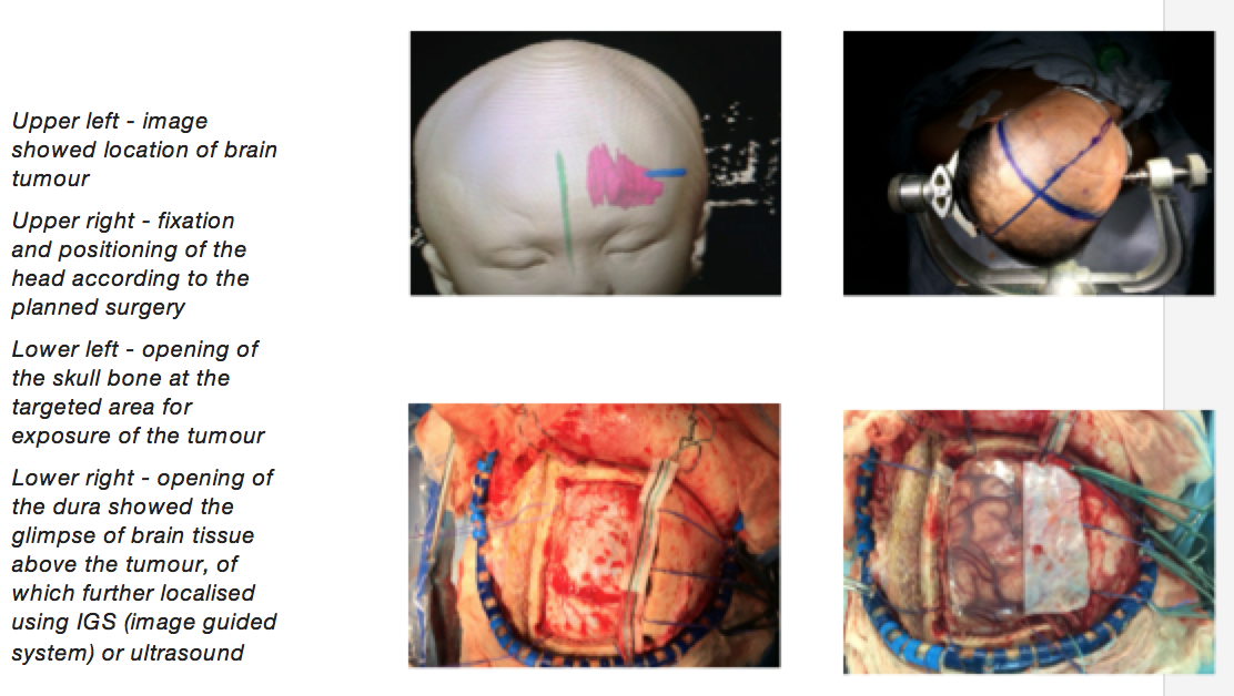
 Hello again readers…Understand the pictures? Have a guess.
Yes…You guessed correctly. These are pictures of a brain tumour in three different views and from the calculations using software available, you get the lower right picture; brain tumour. It is located slightly over to the right of the head, and inside of course. The purple colour is to highlight the tumour, of which I identified and coloured myself, in order to help patient and relatives understand how significant the tumour is, in term of size and location. Recognition of a problem in the brain using these complicated images needs proper training and understanding on how the pictures are being taken.
Reading radiographic imaging is not easy. Sounds easy? Look easy? It is a language of its own and you need to train personnel to read them in a correct manner. I am sure if I don’t colour the tumour, you will not be able to recognise it. Especially when you have three different views of the same thing and what the brain does not know, the eyes will not see until directed to the particular problem.
These advanced imaging acquisitions are available in our country. You do not have to go overseas to get these sort of images. In addition, our country has neurosurgeons who are trained to read such images. They are trained, including me, so that we can explain to you and plan the surgery, if needed.
You might be wondering why I started this article by showing you pictures as above, and of course, more to come. The above shown picture arises from series of imaging taken in the radiology department. The raw data from these images will then be used in a software machine to calculate and generate understandable pictures. This complex process comprises the Image Guided System (IGS) of which many planned surgery will be used as an adjunct to us, the neurosurgeons, in order to have safe surgery.
When you really understand the complexity of the acquisition of the image and comprehension needed by a neurosurgeon before he, or I in that matter, embarked on the treatment of the disease, namely brain tumour, you would understand that it is a difficult matter before making any decision. Especially the decision by you, the patient in regards to the treatment that I would offer.
“Brain tumour and need an operation. I came to see you about something else, doctor. My vision is blurred”. Or, “my foot is weak and I am having difficulty in doing the things I like to do!. And now you are telling me that I have a brain tumour?!. Wow.. I cannot believe my luck, really.”
I understand the conflict that you have. I understand the difficulty in grasping the concept of the brain tumour can cause any of your problems. I do understand the hesitation to accept the treatment offered by me, or any of my colleagues, the neurosurgeons, to you, the patient. I do understand that you are afraid. In fact, I could not understand if a patient of mine did not feel afraid about having a surgery, especially their brain.
Who is not afraid of getting a surgery done on them?
Why me?
Why do I have a tumour in my brain?
Do I need the brain surgery?
These are some questions that the patient, including the relatives have when they see me, a neurosurgeon. I don’t have all the answers but I hope by explaining with pictures and model, I can explain clearly to you that as a neurosurgeon, we have also felt the same. The only difference is that we are trained what to do in such cases. We are trained to take out the tumour in the brain. We are trained to manage this disease process.
 The next picture that I would like to show you is quite similar to the initial picture, except that the side is different. As mentioned in the legend of the pictures, they were not related to each other but will illustrate the complexity of taking out the brain tumour. Planning the surgery always depends on the location of the tumour. With or without IGS, I would locate the tumour using the available imaging and plan for further imaging in order to make a plan about the surgery and to make the surgery safer. As you can see, planning is important before we do our
job as neurosurgeons.
You may say that the subsequent picture looks painful – 3 sharp pins on the head just to fix into position? Is it really necessary?
The usage of fixation method is necessary so that we can work in a precise manner, meaning that our working nature can be down to millimetre of dissection and visualisation. So you can understand why neurosurgeons are so meticulous by nature. The area of surgery is so sensitive that we have to be such a fussy person in regards to taking out the brain tumour. And it is for the sake of the patient, you.
After the planning, there is an execution of the plan. Surgery is not complete without doing the surgery and the last two pictures will illustrate that point. I need to have an access to the tumour by opening the skull in a targeted manner, and later opening of the dura. You might be asking what do the pictures show? The left bottom picture shows an opened skull bone after skin and flap being reflected to expose the site of the surgery. Using specific instruments to cut the bone in such manner, I managed to expose the part of the brain that will lead me to the tumour. The not so clear whitish structure under the bone before the brain matter being exposed is called dura. Dura is a layer of covering the brain that needs to be opened before reaching the target. As you can see from the last picture (right bottom) clearly showed brain matter with vessels in the surgical field after reflecting the dura to the opposite site.
With such exposure, a tumour that was shown to the patient and relatives beforehand during the planning stage, is taken out. Reconstruction and restoration of normal appearance is done once the surgeon is happy with the tumour excision.
So as you can see from the description above, a neurosurgeon does his or her work in a meticulous manner. We understand that each and every one of our patients have a lot of worries once they were told that they have brain a tumour and surgery is deemed necessary as part of the treatment. We can have the treatment here as more and more Malaysians are capable of fulfilling the task as neurosurgeons. Gone are the days that we need to go to other countries in order to receive such treatments. Gone are the days that we depend on others in order to treat brain tumors. Leave the difficulties in planning and doing the surgery to us, the Malaysian neurosurgeons.
Dr. Rahmat is a neurosurgeon who believes strongly that knowledge empowers the public to make the right choices. Read more about him at The Team page.
[This article belongs to The Malaysian Medical Gazette. Any republication (online or offline) without written permission from The Malaysian Medical Gazette is prohibited.]
