Source: wtlab.iis.u-tokyo.ac.jp
Introduction
So what is an echocardiogram?
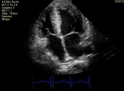
To the general public, this form of examination is simply put, a non-invasive medical procedure that allows the doctors or the cardiac technicians to look into your heart in a real time imaging motion. They could then capture pictures of your heart in certain angles and record a video of it delineating the movement of your valves and heart walls, detailing its chamber size and its volume, its predicted mass and pump efficacy and an assortment of other information deemed necessary. It is ultimately a harmless procedure as it utilizes ultrasonography technology through the usage of a probe that generates echo beams into the internal organs. The organs then reverberates the sound that hit them and these echo back towards the probe transmitting live – black and white imagery on the screen attached to the machine.
Indications – Why You Need an Echo
Please note that these are only SOME of the indications for an echo.
- To look at the heart’s pumping mechanism and its efficiency
- To look at your heart valves allowing them to detail any abnormalities
- To look at the wall motion of the heart as in patients with heart attack or a history of one such attack would lead to weakening of the said area of the wall
- To look at the thickness of the heart walls
- To look at the chambers of your heart to which there are four – a swelling of these chambers may indicate a form of heart failure
- To look for fluid surrounding the layers of the heart (pericardial effusion)
- To look for abnormal growth within the heart

Source: www.havi-north.com
Preparation
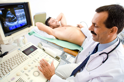
As this is a rather simple and quick procedure, there need not be any form of drastic preparation in detail. Gel will be applied to your chest to improve echo transmission by the probe. Several sticker leads – will be attached onto your chest to record the activity of your heart and this helps the technician/doctor to identify the phases of your heart pumping activity. You do not need to fast beforehand. A technician will position you on an attached bed next to the machine and help you to turn to your left where most of the imaging of the heart would be done. Some pressure would be elicited at some point should your heart becomes difficult to image, and this may cause some slight discomfort from the probe. Quite a number of positioning of the probes would be expected, as the technician/doctor would want to capture the heart’s image at differing angle allowing him to get a full detail of your heart and locate any significant abnormalities.
Results
You should expect immediate analysis and interpretation of your echocardiogram report. Echo findings will be interpreted by your doctor.
The Future
With the advancement of imaging technology and science of echocardiographic study, more and more information are made available through the simple procedure of echocardiography. With more money spent on the revolution and research of echocardiographic machines, the more common 2-D echocardiography which produces black and white imaging are now being replaced by 3-D and 4-D echocardiography that reveals colorful images alongside the capability to alter and slice through the heart images depending on the needs and wants of the doctors and the technicians doing it.
Final Words
Echocardiography complements the array of cardiac investigations made readily available for both the general physicians and the cardiologists alike, as it is not only safe, but also cheap. Results are available rapidly and interpretation doesn’t usually take up a lot of time. It aids both in the outpatient consults and as well as the inpatient ones to differentiate the subtle clinical signs of a possible heart attack. Echo is not the gold standard of diagnosing a new onset of a heart attack or an ongoing one. It merely complements one’s clinical assessment of the patient alongside electrocardiography (ECG) and blood results. Ultimately, it is still a doctor’s job to collate all the information available and to interpret them soundly.
Dr. Chiam Keng Hoong is an internal medicine physician and a MRCP holder. He currently works in Sabah.
[This article belongs to The Malaysian Medical Gazette. Any republication (online or offline) without written permission from The Malaysian Medical Gazette is prohibited.]
