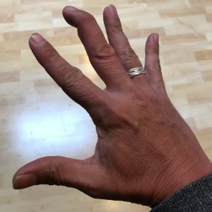
 As the name suggests, mallet finger refers to the condition where the finger is deformed to look like a mallet, an instrument which is used to hit things, somewhat like a hammer. If you are an unfortunate patron at a fine dining restaurant serving great chilli crabs, you may have seen a tool given with the crab dish to knock open the hard shell of the crab, and this is called a mallet. I say unfortunate because it really is much easier to eat crabs with your bare hands than to use a mallet, and many diners would agree with me on this!
A mallet finger deformity usually occurs following trauma. The common cause is due to a direct blow on the tip of a fully extended finger, forcing the tip to bend. For this reason, the most affected fingers usually are the more prominent stand out ones, this includes the index finger, the middle finger (the longest in the finger family), the ring finger and occasionally the little finger as it is more exposed at the sides of the hand; in other words – the thumb is least affected. Occasionally a laceration wound (or a cut) over the dorsal (back) aspect of the finger may result in the same deformity.
A mallet finger is a result of the extensor tendon over the distal interphalangeal joint (DIPJ) of the finger being injured or cut, occasionally the torn tendon pulls off a piece of the distal bone of the finger resulting in an avulsion fracture, hence the term soft tissue mallet injury (injury to the extensor tendon only) or bony mallet injury (extensor tendon injury with avulsion fracture of the distal phalanx).
Symptoms usually are pain and deformity over the distal phalanx, following trauma often after a sports injury. Physical examination would reveal a flexion deformity of the finger at rest, and there is reduced or absent active extension over the DIPJ i.e. the fingertip bends at rest with inability to straighten it actively. A plain radiograph, or an X-ray is often done to exclude any bony involvement, or an avulsion fracture, and this is the only investigation needed for this condition.
A soft tissue mallet injury occurring for a duration of less than 12 weeks requires only splinting. A conservative approach is also indicated if the bony fragment seen on x-rays are too small or not displaced. Splinting is done over the affected finger using a figure of 8 splint to immobilize the DIPJ, and keeping the proximal joint of the finger (PIPJ) free to move. Immobilization is necessary for a duration of 6 weeks.
Surgery on the other hand (puns intended!) is indicated if the bony fragment is large (more than 50% of the articular surface), in chronic injuries, or when conservative treatment fails. Surgical options include using temporary K-wires driven into the finger, tendon reconstruction which may include suturing from the skin into the extensor tendon without a surgical incision – also known as tenodermodesis, or even fusing the finger together. Fusion or arthrodesis of the DIPJ (distal interphalangeal joint) of the affected is done only as a last resort when all other modalities fail, in a painful arthritic joint.
Dr Mohamed Faizal bin Hj Sikkandar
Consultant Orthopedic Surgeon and Senior Lecturer
UiTM Specialist Center, Sungai Buloh Campus
Special Interest in Hand and Microsurgery
